Background
HHT is an under-diagnosed hereditary disease which predisposes patients to developing abnormal blood vessels called telangiectasias. These are abnormal blood vessels because they represent direct connections between arteries and veins; normally capillaries are located in between arteries and veins. These abnormal blood vessels can involve small blood vessels near the surface of the skin or larger vessels within internal organs such as the lungs. When they involve the lungs, they are called pulmonary arteriovenous malformations (PAVMs). A PAVM can involve a single artery draining into a single vein or multiple arteries draining into multiple veins.
Many patients with HHT are unaware that they even have this disease because they do not have obvious symptoms. The most common symptom for a patient with HHT is recurrent nosebleeds. This is due to the presence of telangiectasias under the lining of the space inside the nose. If these telangiectasis occur in the stomach or intestines, they can result in GI bleeding, which can result in anemia. Pulmonary AVMs are often asymptomatic, but can cause patients to have shortness of breath or poor exercise tolerance. They can also result in a stroke or brain infection because clot or bacteria can pass through the lungs directly into the brain. Congestive heart failure is another potential problem associated with an AVM.
Procedural Details
At Albany IR, we offer embolization procedures to treat patients with PAVMs. This is an outpatient procedure that involves the placement of devices within the artery or arteries supplying the PAVM in order to stop the flow of blood passing directly into the venous system. A successful embolization procedure significantly reduces the risk of the above-described complications and helps improve exercise tolerance and shortness of breath.
The procedure is performed through a small catheter that is usually introduced into the common femoral vein near the right hip. Using x-rays for guidance, this catheter is advanced into the inferior vena cava (the main vein draining blood from both legs) and then into the heart. From the heart, the catheter can be advanced into the pulmonary artery bringing blood into the affected lung. At this point, an angiogram is performed, which involves injecting contrast material into the catheter so that pictures of the arteries can be obtained. These pictures can help determine the exact location of the PAVM and can identify which artery or arteries will need to be embolized in order to completely treat the PAVM. The catheter is then advanced directly into the affected artery or arteries and embolization is performed. The devices used for this procedure, such as plugs and detachable coils, have greatly increased the safety of the procedure because they are not released into the circulation until they are appropriately placed.
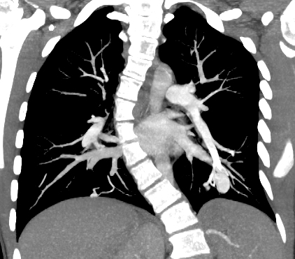
This is a coronal view from a CT angiogram that demonstrates a pulmonary AVM arising from the lower lobe artery of the left pulmonary artery.

This is an image from a pulmonary angiogram showing the PAVM in the left lower lobe before embolization.
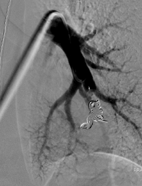
This is an image from a pulmonary angiogram after embolization of the left lower lobe PAVM.
Results
Following embolization of a PAVM,